We began our third burial today. In case you are wondering, after we receive our burial, we carefully remove the pieces from their bag(s) and begin to lay them out in anatomical position on our workbench. This can be a long, complicated process when there are many fragments, especially if their is co-mingling. (Our second burial had 103 unidentified bone fragments and the cranium was in numerous pieces, plus a co-mingled maxilla.)

Once they are all laid out, we fill out some forms. The first is a skeletal inventory form. It has sections for each area of the skeleton (cranial bones, axial bones, appendicular, long etc.) We shade a diagram to show which bones are present. We give the bones a F (1-25%), P (25-75%) or C (75-100%) based on how fragmentary, partial or complete they are. The next form is used to assess the level of fusion of the ephiphyseal plates (mandible, long bones, ilium etc). We choose 0 for open, 1 for fusing and 2 for fused. Assessing fusion is helpful in age estimation.

This iliac crest is open.

The humerus has a proximal and distal epiphyseal plate. They are both open.

Next, we measure for age estimation. (Broken or incomplete bones are not measured.) There are metric charts to help interpret this data.

Tooth sorting! We differentiate between deciduous and permanent, maxillary and mandibular, right and left. This is a mix of deciduous and permanent. Oftentimes, the teeth are no longer in their respective arcade. We shade them in on a diagram and then estimate age. We use standards from Ubelaker and the London Tooth Atlas to do this.

We write up general comments next and are ready for peer-review. We peer-review another group's work too. Once the peer-review is complete, we move the skeleton to a black cloth for photos.
Our second burial laid out for photos. (see the fragments along the bottom right!)

Lastly, we carefully pack the bones back into their respective bags and return them to the director with our paperwork.
Other photos from today:
This is a C2 (second cervical vertebra) or axis. C1, also called the atlas, sits on top of C2 and this is what allows you to rotate your head. I think it looks like a guy flexing his arms. The dens is the part that sticks out from the top.

The thunder rolls...
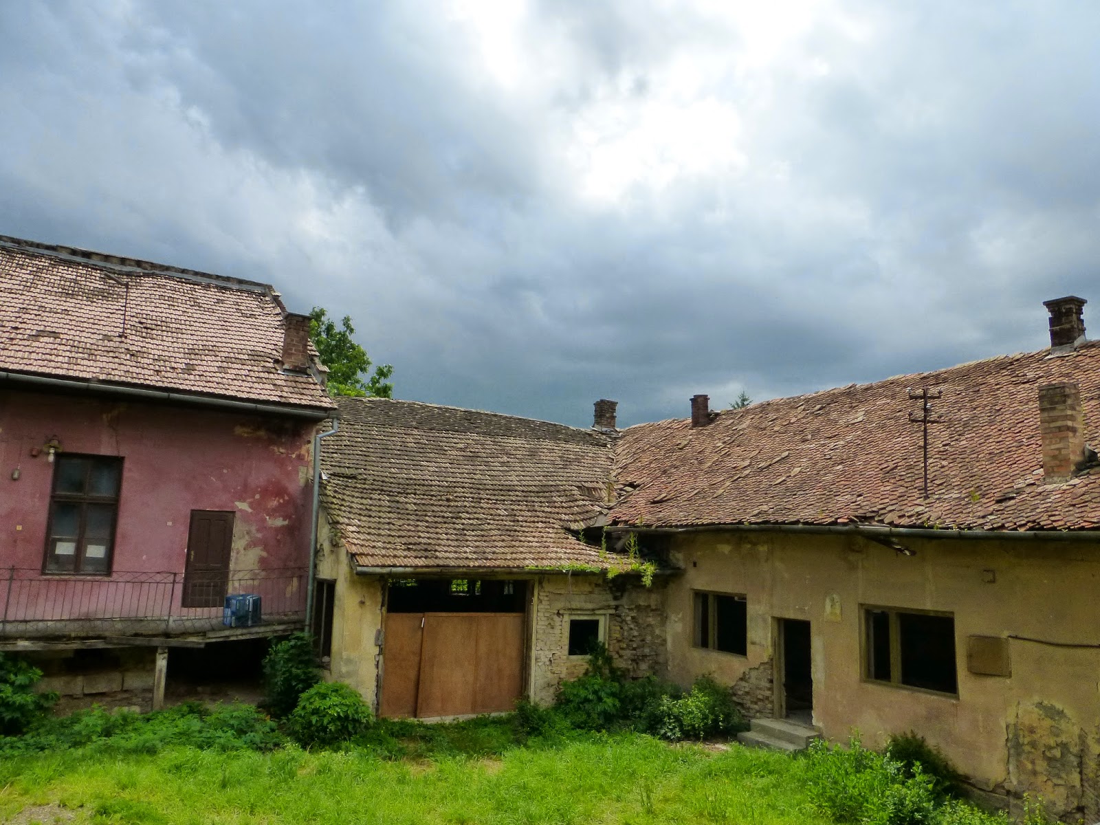
Rainy afternoon...

Lola gazes pensively out the lab window. I think she might need a Gelato break.

It's open lab tonight. We have two more bone quizzes to go, plus an annotated bibliography with 10 sources, a couple of lab assignments and a presentation left with just over a week to go.
I didn't photograph them, but a Hungarian news team filmed us in the lab today. They also interviewed our director and one of my classmates. I will post a link for that when it is up.
No comments:
Post a Comment